Tetrachromacy: who has a fourth cone, and why no online test can find it
Most people see color with three types of cone, the light-sensitive cells in the retina that respond to color. Some women have a fourth type, and a very few of them seem to use it to tell apart colors that look identical to everyone else. Here is what the studies actually found, which popular numbers hold up, and a calculation showing why a test on your phone or monitor cannot detect it.
Short answer
- Tetrachromacy means seeing color with four kinds of cone instead of three. In people it is possible only in some women whose fathers or sons have a mild form of red-green color blindness.
- About 12% of women are such carriers, according to the main study on the subject. Having four cone types does not by itself add a dimension of color.
- In that study (Jordan and colleagues, 2010) 1 of 24 carriers passed every test for four-dimensional color vision. Nobody has measured how common that is in the population.
- If your son has deuteranomaly, the most common mild form of red-green color blindness, you belong to the group behind that result: all 24 women were found through a color-anomalous child. See what the result means for you.
- No online test can tell you. A screen builds every color from three lights, so any two colors that look the same to a three-cone eye are also the same for a fourth cone. The lab tests use pure spectral lights: light of a single wavelength, like a laser pointer's, which a screen cannot produce.
Key numbers
- Some 12% of women are carriers of the genes for anomalous trichromacy, a mild color deficiency, so their retinas should contain four cone types (Jordan, Deeb, Bosten and Mollon, 2010).
- In that 2010 study, 1 of 24 carriers of deuteranomaly passed every test for four-dimensional color vision.
- Anomalous trichromacy, the mild form of red-green color blindness, affects 6% of men: deuteranomaly 5% and protanomaly 1% (Jordan and colleagues, 2010).
- In our model of the one carrier who passed (cone peaks 426.3, 530, 545 and 557 nm), her fourth cone catches 17% less light from a red-green mixture than from the 590 nm orange it matches; on our model three-primary screen the two copies differ for it by only 0.001%.
- The popular claim that tetrachromats see 100 million colors rests on arithmetic (100⁴), not on a measurement; we found no study behind it.
What tetrachromacy is
Each cone type responds to a broad band of wavelengths, measured in nanometers (nm), the unit of wavelength. The three types are named for where they respond best: S for short wavelengths, M for medium and L for long. With three types, any light, however complex its spectrum, comes down to three numbers, and two lights with the same three numbers look the same. Color scientists call such pairs metamers. A screen works by making metamers: its mix of red, green and blue only has to give the same three numbers as the color it copies. A tetrachromat would have a fourth independent number, so some metamers would look different to her.
Gabriele Jordan and John Mollon separated two forms in 1993. In weak tetrachromacy a woman has four cone types, but her nervous system keeps only three independent signals: she accepts the same color matches as everyone else, though her matches may shift when the surroundings change. Strong tetrachromacy means the brain gets four independent signals, and she needs four variables to match all colors. Put simply: in the weak form the fourth cone is there but makes no visible difference; in the strong form she sees color differences other people cannot. The popular idea of a tetrachromat who sees extra colors is the strong form.
Who could have a fourth cone
In anomalous trichromacy, the mild form of red-green color blindness, one cone pigment is shifted along the spectrum. Jordan and colleagues give 6% of men for it: deuteranomaly, where the green-sensitive (M) pigment is replaced by one closer to the red-sensitive (L), in 5%, and protanomaly in 1%. The genes are on the X chromosome. The mother or daughter of such a man can carry the normal genes on one X and the shifted one on the other, and since only one X works in each cone cell, her retina should contain four cone types.
How many women is that? The 2010 paper opens with “some 12% of women” as carriers of anomalous trichromacy, and in its discussion puts carriers of deuteranomaly alone at about 10%, citing a 1979 book by Pokorny and colleagues. The 1993 paper gives about 15% as carriers of any X-linked color deficiency, a wider group that includes carriers of full red-green color blindness. Its authors expected strong tetrachromacy “only in carriers for anomalous trichromacy,” the mild form. So the 12% and the 15% describe different groups, and neither counts tetrachromats.
How few use it: the studies
Jordan and Mollon (1993) tested 43 women, 31 of them known carriers. Most carriers of anomalous trichromacy showed no sign of strong tetrachromacy. Eight of 14 could not find a red-green mixture that matched a yellow light on a large field, something no other subject did. That shows their color matches differ from normal; on its own it does not show a fourth dimension of color, which is what the next task looked for. One carrier, labeled cDA7, set unusually consistent matches in a task designed to reveal a fourth dimension, and the authors kept her as a candidate.
Jordan, Deeb, Bosten and Mollon (2010) then tested 24 carriers of deuteranomaly, each with at least one color-anomalous child, plus 7 carriers of protanomaly and two control groups. In the main test the woman looked into an optical instrument at three flashes: two of pure orange light at 590 nm and one mixture of red (670 nm) and green (546 nm) light. For a normal eye some mixtures look exactly like the orange. Her task was to pick the flash that looked different. Most carriers made errors in the same region as people with normal vision. One, coded cDa29, made no errors at any ratio of red to green: whatever the mix, she could always pick the odd one out.
She also stood out in the second test, in which participants rated how different pairs of painted discs looked. Her ratings showed a color dimension that her two sons with mild red-green color blindness (deuteranomaly) share and her son with normal color vision lacks. Sequencing showed normal M and L genes plus a hybrid, and the authors estimate the three pigments peak at about 530, 545 and 557 nm. A different set of laboratory measurements gives 532, 551 and 563 nm. Discover magazine described her in 2012 as a doctor living in northern England.
The authors are careful about what one case means. They write that most carriers do not show four-dimensional color vision, and that three well-separated pigments may not be enough on their own: one carrier with an 11 nm gap between pigments showed nothing unusual. The 24 women were found through their color-anomalous children, so 1 in 24 is a result for this group and says little about women in general.
If your son has the mild form of color blindness, this group is you. Jordan and colleagues name the mother (or daughter) of a man with the mild form, anomalous trichromacy, as a candidate for tetrachromacy, and they recruited the 24 women as mothers of color-anomalous children, usually sons. For a mother of a son with deuteranomaly, 1 in 24 is the closest estimate there is, from a small sample. The result covers only the mild form: the study tested only carriers of deuteranomaly and protanomaly. If your son's diagnosis says deuteranomaly or protanomaly, that is the mild form.
Can an online test tell if I'm a tetrachromat?
No, and for the same reason a screen cannot show colors that only a tetrachromat would see. Every pixel on a phone, laptop or TV is a mix of three lights: red, green and blue. A screen can vary only how much of each it sends, so all the colors it can make form a three-dimensional family. A fourth cone looking at that family learns nothing new. Its response to any screen color is a fixed blend of the responses of the other three cones. Two colors that a normal eye sees as identical are, on a screen, the same three numbers, so they are identical for the fourth cone as well.
Quizzes that show bands of similar colors and ask how many you can count measure something else: your screen, your lighting, your attention, and how you count. The lab tests above use pure spectral lights or physical paints, because those can differ in ways a screen cannot reproduce. Jordan's group at Newcastle University makes the same point in its FAQ: screens do not give enough color information, so an online test cannot investigate tetrachromacy.
The calculation
We took the four pigments estimated for cDa29 (S at 426.3 nm, M at 530, the hybrid at 545 and L at 557) and drew their absorption curves with the standard template we use on our animal vision pages.
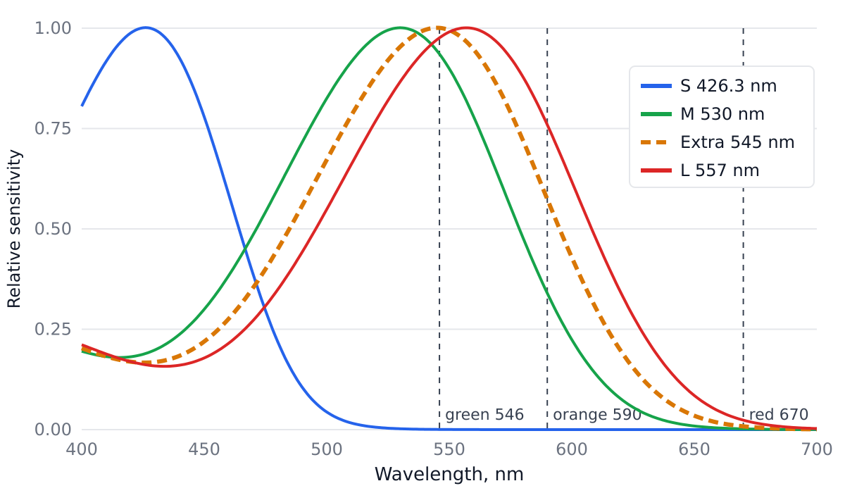
Think of each cone as a light meter: how much light it catches is its reading. First the real-world pair. We mixed 670 nm and 546 nm light until the M and L cones caught exactly as much from the mixture as from 590 nm orange. The S cone catches almost nothing from either (0.02% of the L catch at most). So for a normal eye the two lights are a perfect match. The extra cone catches 17% less from the mixture than from the orange. With the alternative peaks (532, 551, 563 nm) the gap is 20%.
Then the screen. We modeled one with three primaries and asked it to copy both lights as closely as a normal eye can judge. Both copies come out as the same red, green and blue settings, and the extra cone catches the same amount from each: the difference is 0.001%, left over from the S cone's trace response. The 17% gap is gone. The copy is not even right for the extra cone: it catches 5% less from the screen orange than from the real one, an error a normal eye never notices.
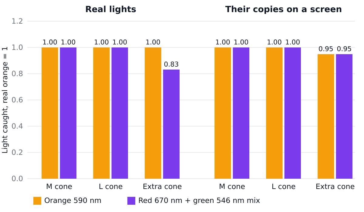
Put plainly, a screen has only three knobs. Whatever you set them to, the extra cone's reading is always about half the L reading plus half the M reading, so it never tells the brain anything the other three cones did not. That fits the first figure, where its curve sits between M and L. As a formula, on this screen the extra cone's response to any color equals 0.47 × L + 0.56 × M − 0.05 × S, exactly. In linear algebra terms, the 4 × 3 matrix that turns red, green and blue settings into four cone responses has rank 3. Our primaries are a model (their shapes are given under “How we computed this”), so we repeated the test on 1,000 screens with random primaries. On every one, the two copies differed for the extra cone by at most about 0.005%. The rank argument holds for any screen with three primaries, however wide its gamut.
How large the real-world gap is depends on where the extra pigment sits. In our model, with M at 530 and L at 557 nm, an extra pigment at 534 nm gives a gap of 8% on this test and one at 553 nm gives 9%; the largest, 17%, is near 544 nm. These are differences in light caught by the cones. Whether a brain turns them into a visible difference is what the lab tests measure, and for 23 of 24 carriers it did not.
One more number puts the screen in context. For pure spectral lights from 400 to 700 nm, the best blend of L, M and S still misses the extra cone's response by 4.0% on average. Smooth surface colors of the kind our paint mixer models, lit by daylight, give a miss of only 0.03%. Those surface spectra are synthetic, so take this as a hint: the extra cone has the most to add with narrow-band light, which is exactly what the lab tests use. A screen leaves it nothing to add.
Popular claims, checked
“12% of women are tetrachromats”
Wrong
Jordan and colleagues write that some 12% of women are carriers of anomalous trichromacy, which means their retinas should hold four cone types. The same paper found the extra dimension of color in 1 of the 24 carriers of deuteranomaly it tested. Having the cones and using them are different things.
“Tetrachromats see 100 million colors”
Untraceable
The figure is arithmetic: if each cone type gives about 100 steps, three cones give 100³ = 1 million and four give 100⁴ = 100 million. Discover wrote in 2012 that “it’s possible these so-called tetrachromats see a hundred million colors” and gave this arithmetic as the reason. We found no measurement behind it, and Kimberly Jameson of UC Irvine, who studies tetrachromacy, writes that there is no scientific basis for it.
“Take this test to find out if you are a tetrachromat”
Impossible on a screen
A screen mixes every color from three fixed lights, so it cannot show the colors a fourth cone would add. The Newcastle team that found cDa29 says in its FAQ that an online test cannot investigate tetrachromacy. Our calculation above shows the reason.
“Men can be tetrachromats too”
Not by this route
The genes for the M and L cone pigments sit on the X chromosome. A man has one X, so he has one version of the array. A woman with two different versions can express both, one in some cones and one in others. That is the route the studies above test.
The 12% figure also travels in this form on the University of Washington ophthalmology site (December 2023), which says about 12% of women “have it”, next to a statement attributed to Jay Neitz that tetrachromats can see 100 million colors. We found no paper giving that number. Jameson traces the idea behind it to an article by Neitz, Carroll and Neitz in Optics & Photonics News (2001), which we could not read in full.
What you can actually find out
- Whether you could be a carrier. The Newcastle team looks at male relatives on the mother's side: if your son, father, maternal uncle or maternal grandfather is very slightly color-blind, you may carry the gene for the mild form. Our color blindness simulator shows roughly how they see.
- Whether you have the genes. The University of Washington page above mentions a genetic test. A gene result shows that the pigment can be there; it cannot show that your brain uses it.
- Whether you use them. That takes a lab with spectral lights, like the anomaloscope and the tests described above. The Newcastle project recruits participants through its site.
Birds have four types of single cone. Our bird vision page shows what a screen can show of that and says what it cannot.
If you design for screens, what you can plan for is red-green color blindness: see the color blindness simulator and colorblind-safe palettes.
Strong tetrachromacy is real but has been confirmed in very few people, and no screen can find it. If you want to know, the route is a carrier check and a lab.
How we computed this
Cone curves: the Govardovskii template for visual pigments without lens or macular pigment, each normalized to its peak, as Jordan and colleagues normalized theirs; pigment density is not modeled. Leaving out the lens does not change the result for pure spectral lights: a filter in front of the retina dims each wavelength equally for every cone, so it changes the amounts of light needed for a match and leaves the gap for the extra cone the same. Model screen: Gaussian primaries peaking at 615, 535, 455 nm, 25, 35, 22 nm wide at half height; the random screens use peaks from 590 to 650, 510 to 560 and 440 to 470 nm and widths from 15 to 60 nm. Pure orange at 590 nm lies a hair outside this screen's range of colors, which does not change the argument. Surface spectra: spectral.js under CIE D65. Computed on November 8, 2026 with our own script.
Sources
- Jordan G., Deeb S. S., Bosten J. M., Mollon J. D. (2010). The dimensionality of color vision in carriers of anomalous trichromacy. Journal of Vision 10(8):12. doi:10.1167/10.8.12 (author PDF; full text read).
- Jordan G., Mollon J. D. (1993). A study of women heterozygous for colour deficiencies. Vision Research 33(11):1495–1508. doi:10.1016/0042-6989(93)90143-K (full text read).
- Newcastle University Tetrachromacy Project, FAQs.
- Greenwood V. (2012). The humans with super human vision. Discover.
- Jameson K. A. (2014). Tetrachromacy myth busting, UC Irvine.