How vision evolved: from a light-sensing cell to the human eye
An eye did not appear in one step. Living things today still carry every stage, from a cell that only knows whether it is light or dark to the camera eye you are reading with. We took one photo of a living room and ran it through a model of each stage, using the angles measured in published studies. The pictures below show roughly what each kind of eye can resolve. The stages are ordered by eye design, each shown by an animal that has it, and most of those animals are not our ancestors. The evolution of vision simulator runs the same stages on your own photo or camera.

Short answer
- Vision built up in four kinds of task, in order: telling light from dark, telling which way the light comes from, seeing a crude image, and seeing a sharp one. That is the scheme of Dan-Eric Nilsson of Lund University, and each step still exists in living animals.
- It was fast in geological terms. Nilsson estimates that the path from the first opsin, the protein family behind animal light sensing, to sharp vision took about 170 million years and was largely complete by the start of the Cambrian, which he puts at about 530 million years ago.
- Our color vision has its own, later story. The ancestors of marsupials and placental mammals lost two of their four cone types, probably during a nocturnal phase; Old World primates gained a third cone back by duplicating a gene.
- The pictures are models. They show how much detail and color each eye design lets through; what a trilobite or a jellyfish experienced is beyond them.
Key numbers
- Dan-Eric Nilsson (2013) estimates that the path from the first opsin to sharp vision took about 170 million years and was largely complete by the start of the Cambrian, which he puts at about 530 million years ago.
- The compound eye of the trilobite Schmidtiellus reetae, more than half a billion years old, has fewer than about 100 ommatidia, each about 10° from its neighbor (Schoenemann and colleagues, 2017).
- Lamb, Collin and Pugh (2007) conclude that a camera-like vertebrate eye must have existed by about 500 million years ago, when lampreys and jawed vertebrates split.
- The ancestors of marsupials and placental mammals lost two of their four cone types, probably during a nocturnal phase about 150–200 million years ago (Hunt and colleagues, 2009).
- Human cone pigments peak at about 419, 531 and 558 nm (Dartnall, Bowmaker and Mollon, 1983).
How the pictures are made
Every stage starts from the same photo. We assume the photo spans 60° from side to side, our own choice, and turn each published angle into pixels on that basis: a 25° blur spans 25/60 of the picture's width. A blur angle is treated as the width of the blur spot at half its height. Stages with no color data are shown in gray, as brightness only.
What the pictures leave out: how bright or noisy each eye's image was, what the animal's nervous system did with it, and any color an extinct animal saw. Opsins, the light-sensitive proteins, do not fossilize.
A timeline of the dates we could check
- About 3.3–3.6 billion years agoCyanobacteria arise (molecular-clock estimates, which disagree). When their light-steering arose is not dated.
- 755–711 million years agoThe first animal opsin appears. It “might not have been light-sensitive.”
- 711–700 million years agoThe main animal opsin families split.
- About 540 million years agoThe Cambrian explosion begins.
- About 530 million years agoBy Nilsson’s estimate, the path to sharp vision is largely complete.
- More than half a billion years agoThe trilobite Schmidtiellus has a compound eye.
- About 515 million years agoAnomalocaris has eyes with at least 16,000 lenses.
- By about 500 million years agoThe vertebrate camera eye exists.
- About 150–200 million years agoEarly mammals go through a nocturnal phase thought to have cut their cone types from four to two.
- Undated in our sourcesOld World primates regain a third cone.
Stage 1: light or dark
The simplest light sensor reports one number: how bright it is around me. Nilsson calls this class I, with a detection angle of 360°. It has no direction and no picture. That is still enough to keep a day-night clock or to sense depth in water.

Some archaea, single-celled microbes, use a light-sensitive protein called sensory rhodopsin I. Spudich and Bogomolni reported in 1984 that it lets the cells “distinguish colour”: orange light attracts them, and when a second photon is absorbed, the signal turns into a repellent (Olson and colleagues, 1995). So the first “color vision” was a choice of which way to swim, with no picture involved. A screen can't show that, so the figure stays one gray value.
Stage 2: which way the light comes from
The next task is direction. The cyanobacterium Synechocystis is a sphere about 3 micrometers across, and the whole cell works as a lens: it focuses an image of the light source on the edge of the cell opposite the source, and the cell moves toward the light. Its authors call spherical cyanobacteria “probably the world's smallest and oldest example of a camera eye.” The angular resolution is about 21°.
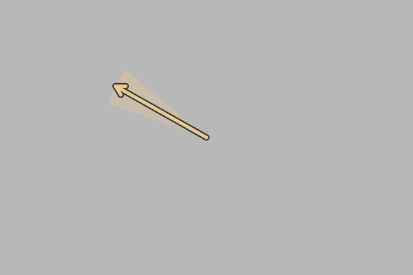
Single-celled algae such as Chlamydomonas solve the same problem differently. A pigmented eyespot shades the photoreceptor from one side, and as the cell rotates, the signal rises and falls, which tells it where the light is.
Among animals, the larva of the marine worm Platynereis has a simple larval eye with a field of about 180° (Nilsson, 2009). Our figure compares the left and right halves of the photo as a stand-in for that kind of direction sensing.
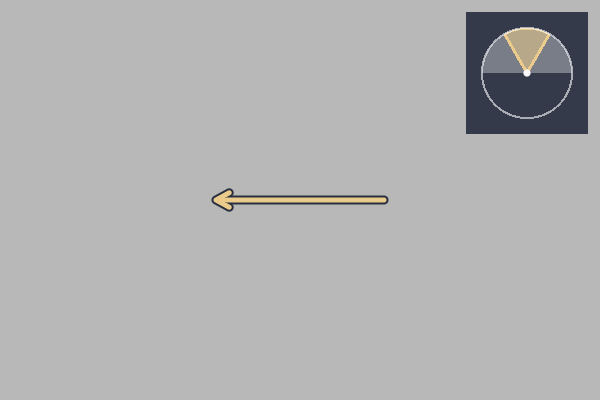
In the figures these first three stages look almost alike, and that is accurate. A cell that only knows brightness and direction has nothing more to show.
Stage 3: a crude image
Put several receptors in a pigment cup and each one sees a different patch of the world. Nilsson gives about 25° per patch for this class III, “low-resolution vision”; a flatworm's eye cup is a living example of the design. That is too coarse for objects, but enough to find a dark shelter or the line of the horizon.

Stage 4: compound eyes in the Cambrian
By the Cambrian, eyes are in the fossil record. One of the oldest belongs to the trilobite Schmidtiellus reetae, more than half a billion years old. Its compound eye has fewer than about 100 units, called ommatidia, each about 10° from its neighbor and collecting light over 20 to 30°. The study notes that it lacks the lenses typical of modern compound eyes.
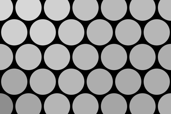
Anomalocaris, about 515 million years ago, had at least 16,000 lenses per eye (Paterson and colleagues, 2011). Its acuity and color vision are not known from the fossils, so we don't draw it.
Stage 5: lens eyes without a brain
Box jellyfish have lens eyes and no centralized brain: their nervous system is a ring nerve plus nerve tissue in the rhopalia, the sensory structures that carry the eyes. The upper lens eye looks up through the water's surface with a field of just under 100°, and its receptors collect light over 10 to 20° (half-width; Bielecki and colleagues, 2013). Garm and colleagues measured responses that fit a single opsin, with its peak in the blue-green, so the animal is color-blind.

Stage 6: a pinhole
The living Nautilus has a pinhole eye: an open cup with a small hole and no lens. A pinhole makes an image but trades sharpness for light. Muntz and Raj measured the smallest angle it can separate as between 5.5 and 11.25°.

Stage 7: the vertebrate camera eye
Lampreys are jawless fish whose eye is close to ours: it has a lens, an iris and muscles to move it. Lamb, Collin and Pugh conclude that a camera-like eye must have existed by the time lampreys and jawed vertebrates split, around 500 million years ago. The southern lamprey Geotria australis has five classes of photoreceptor, all cone-like, and five classes of opsin related to those of jawed vertebrates. Northern lampreys have only two photoreceptor classes.

We know of no measured cone peaks for the lamprey, so we can't compute its colors. The figure keeps the photo's own colors, the way a human sees them, and its sharpness is Nilsson's class IV value for sharp vision (5° in his relaxed limit). The lamprey's own acuity is not in our sources.
Stage 8: color lost and found
Marsupials and placental mammals have two cone opsin classes where their ancestors had four. A review by Hunt and colleagues links the loss to a nocturnal phase “thought to mark the early evolution of the mammals around 150–200 Ma”; the SWS2 and RH2 genes stopped working. Reconstructions cited by Yokoyama and colleagues put the mammalian ancestor's two remaining cone pigments at about 360 nm, in the ultraviolet, and about 560 nm.
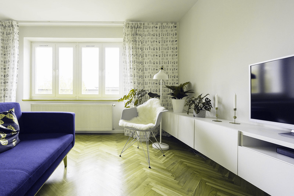
A photo records no ultraviolet, so the 360 nm cone only catches the violet end of what the camera saw. Living mammals have spread out since: Hunt's review gives short-wave peaks from 360 nm in the mouse to 440 nm in the tree squirrel.
Old World primates got a third cone when the long-wave opsin gene was duplicated (Carvalho and colleagues, 2017). Dartnall, Bowmaker and Mollon measured human cone peaks at 419, 531 and 558 nm. That is the photo as you see it at the top of the page.
How fast can an eye evolve?
In 1994 Nilsson and Pelger modeled a flat patch of light-sensitive cells turning into a lens eye through small improvements, each of them useful. With cautious assumptions, they found that it would take “only a few hundred thousand years.” That is the optics of one eye, starting from a patch of light-sensitive cells; the 170 million years in the short answer run from the first opsin. Many articles quote an exact count of generations from the paper. We have only been able to read its abstract, so we don't repeat it.
Nilsson's later review adds that directional light sensing arose independently at least twice before the common ancestor of bilateral animals, and went on to true vision several times in different groups. So the stages above are a ladder of designs drawn from different branches of life. A box jellyfish is not our ancestor, and the stages are ordered by eye type.
What the pictures can't tell you
- The colors of extinct animals. No Cambrian palette is shown.
- How bright or noisy each image was, or how fast it updated.
- What any animal experienced.
- What the single-celled warnowiids do with their “ocelloid,” a structure with analogues of a cornea, lens and retina inside one cell. Its function is unknown, so any picture would be invented.
Try it on your own photo
The evolution of vision simulator runs the same stages on your own photo, a video or your phone's camera, in the browser.
How we made the figures
Sample photo at 600 × 400 pixels, 60° wide. Brightness is averaged in linear light with the Rec. 709 weights; blur is a Gaussian whose width at half height equals the published angle. The early mammal uses a dichromat matrix derived from the two cone peaks with a lens-free photopigment template, D65 light and the Viénot projection, the method of our animal vision pages. Computed on 8 November 2026 with the same stage parameters as the simulator, also averaging light in linear RGB; the simulator draws the blur on a coarser grid to keep the camera fast, so its pictures can differ slightly from these.